Jun 27, 2022

Anterior cruciate ligament: functions and pathologies

June 27, 2022 0 Comments

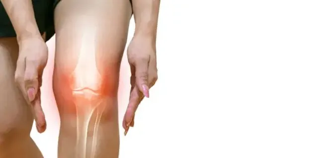
The anterior cruciate ligament (ACL) is located in the middle of the knee (it is part of the "central pivot"). Placed in the indentation of the femur, a real cavity in the middle of the knee, it is oblique upwards, backward, and outwards. 

anterior cruciate ligament,anterior cruciate ligament injury,anterior cruciate ligament tear,anterior cruciate ligament (anatomical structure),anterior cruciate ligament injury (disease or medical condition),anterior cruciate ligament anatomy,anterior cruciate ligament test,posterior cruciate ligament,anterior cruciate ligament function,#anterior cruciate ligament function,#anterior cruciate ligament origin and insertion,anterior cruciate ligament reconstruction


The knee joint is the largest of all. It is divided into three joints:


  • the intermediate patellofemoral joint, between the patella and the patellar surface of the femur (thigh bone);
  • the external femoral-tibial joint, between the external femoral condyle, the external meniscus, and the external tuberosity of the tibia (leg bone);
  • the medial femoral-tibial joint, between the medial femoral condyle, the medial meniscus, and the medial tuberosity of the tibia.

The patellofemoral joint is flat, while the two femorotibial joints are hinged.

Among the various anatomical structures that make up the knee joint are the two cruciate ligaments, which intersect symmetrically in the center of the knee:

  • the anterior cruciate ligament begins at the front end of the tibia, crosses the knee joint diagonally backward, and then attaches to the posterior part of the lateral femoral condyle;
  • The posterior cruciate ligament extends from the rear end of the tibia to cross forward and inward to the anterior part of the medial femoral condyle.

This crossing is accentuated in particular during flexion and extension movements of the knee, or during rotation movements of the leg.


1. What is the purpose of the anterior cruciate ligament?

The two cruciate ligaments are essential for stabilizing the knee. The anterior cruciate ligament controls changes in direction such as rotation and twisting movements. The role of the anterior cruciate ligament is essential: it prevents the tibia from moving too much in relation to the femur and it also prevents excessive rotation of the tibia in relation to the femur.

In approximately 70% of serious knee injuries, the anterior cruciate ligament is stretched or torn.


2. What are the pathologies related to the anterior cruciate ligament?

The main damage to the anterior cruciate ligament is ligament rupture.


The rupture of the anterior cruciate ligament of the knee refers to a retraction of this ligament at both ends. This tear, partial or total, is most often of traumatic origin. It is quite frequent in sportsmen, the main sports at risk being soccer, skiing or even combat sports. These sports put a lot of strain on the knee, with rotational movements that are more likely to cause a ligament tear. The trauma can be direct, with a blow to the knee, for example, or indirect if it is a twisting movement leading to a sudden rupture of the ligament (the foot is blocked on the ground, but the knee pivots).


Thus, the most effective means of prevention to reduce the risk of tearing is muscle strengthening and warming up before physical effort. These conditions will make the knee more resistant during jumps or pivots.


The first symptoms of an ACL rupture appear immediately after the trauma. The main symptoms are severe pain in the knee, local swelling, cracking, a feeling of instability and fragility, as well as problems with the function of the knee (difficulty in stretching the knee, walking, etc.).


Subsequently, the rupture of the anterior cruciate ligament results in instability of the knee and a feeling of discomfort in everyday life. Certain knee movements, such as twisting and turning, are difficult, which can be disabling and affect quality of life. Similarly, when the ligament is damaged, the patient is hampered in his or her sports activities, especially in any activity requiring specific movements such as pivots.


3. What are the treatments?

The rupture of the anterior cruciate ligament cannot heal naturally and may require knee surgery.

The operation, called ligamentoplasty, consists in replacing the ruptured ligament. The principle is therefore a reconstruction of the ligament by autograft (harvesting from the patient himself).

This operation is performed under local or general anesthesia. It is performed under arthroscopy, i.e. with the help of an arthroscope introduced into the joint with a small camera. This makes it possible to visualize the lesions and to operate without opening the knee joint.

Several small instruments are then introduced to perform the surgical procedure.

To replace the ruptured ligament, several types of transplants can be used (patellar tendon, hamstring tendons, etc.).

A short incision is made to remove part of the tendon in question, which will be placed in the knee to replace the ruptured ligament.

The consequences on the tendon from which a part has been removed are minimal, if any because it will heal well and should hardly lose any function.

Post-operative rehabilitation can be done with the help of a physiotherapist or in a rehabilitation center. The goal is to reduce pain, maintain flexibility and mobility of the joint in the first instance, and then recover the muscles in the second instance.

The patient will also wear a splint for several weeks to help support the joint, as well as canes or crutches to relieve the weight of the knee.

Recovery of mobility and muscle strength takes place within a few months. However, a new rupture can always occur in the replacement ligament. Therefore, the state of the muscles is a major element to consider before pushing the knee to its limit, especially in sports. The patient must remain vigilant in his or her sports activities and, in particular, in any sport where the knee tends to rotate.

The results of this technique are nevertheless generally satisfactory. It also allows for the preservation of the rest of the knee structure (meniscus, cartilage, etc.) which degrades less significantly on a stable knee.


4. Who are the anterior cruciate ligament specialists?

Doctors specializing in anterior cruciate ligaments are orthopedic surgeons.

In case of pathology related to the anterior cruciate ligament, the attending physician will refer the patient to a specialist, who will determine the severity of the pathology and suggest or not medical-surgical management.


5. What are the diagnostic and complementary examinations?

The clinical diagnosis to confirm or not the rupture of the anterior cruciate ligament is made by an orthopedist.

The clinical examination of an ACL rupture consists first of an interrogation of the patient to determine the circumstances of the occurrence and to evaluate the symptoms felt. In most cases, at the very moment of the trauma causing the rupture, it is possible to hear a cracking sound in the knee. The patient usually experiences severe knee pain and swelling almost immediately. This is compounded by the instability of the knee, making it extremely difficult and painful for the patient to walk, who may feel as though they are walking into a hole or losing control of their knee. In some of the more severe cases, bleeding may occur as well as a total locking of the knee due to trapped tissue.

However, in some cases of partial rupture, the characteristic cracking sound may not be heard and the patient may still be able to walk.

To confirm the diagnosis, the orthopedic physician then performs tests such as the Lachmann maneuver (or anterior drawer maneuver). The goal is to look for abnormal slippage of the tibia in relation to the femur. To do this, the doctor positions the patient's knee between 0 and 30° in order to put tension on the anterior cruciate ligament bundles. In the case of a healthy ligament, the maneuver is characterized by an early hard stop of the tibia. In contrast, in the case of a rupture of the anterior cruciate ligament, the maneuver is characterized by a less clear-cut and delayed arrest. The reaction of the knee can be compared to the other knee if it is healthy, to confirm damage to the anterior cruciate ligament. In addition, the doctor can look for other possible injuries on clinical examination, such as damage to the menisci or collateral ligaments.

However, due to the swelling and severe pain associated with this method, it is difficult to perform and interpret immediately after the ligament rupture.

When the clinical examination is not conclusive, additional radiological examinations are necessary to characterize the pathology.

An X-ray may or may not reveal the presence of a fracture following the trauma. MRI (magnetic resonance imaging) allows for a very precise diagnosis and observation of the inside of the knee, but it does not allow for the evaluation of the functionality of the remaining ligament, which only a clinical examination can reveal. Therefore, there is no urgency to perform an MRI, which can be performed 1 month after the rupture, except in rare cases. Here again, it is the performance of a good clinical examination that will indicate the need to perform an emergency MRI.

If MRI is not indicated, the doctor may use a more invasive arthroscanner. For this, a product is injected directly into the knee joint to allow visualization of the various tissues. Combined with standard X-rays, these imaging examinations can also identify other lesions such as meniscus damage.


Sources :

Opération ligamentaire

Définition rupture ligaments croisés - symptomatologie

Ligamentoplastie

Principes d’anatomie et de physiologie (livre J Tortora et NP Anagnostakos)

Cerebrospinal fluid: definition, role and associated diseases

June 27, 2022 0 Comments

 Cerebrospinal fluid is a biological fluid in which the brain and spinal cord bathe. Zoom on its functions and on the diseases that can reveal its analysis.

cerebrospinal fluid,cerebrospinal fluid (biofluid),cerebrospinal fluid function,cerebrospinal fluid circulation,composition of cerebrospinal fluid in hindi,cerebrospinal fluid composition,production of cerebrospinal fluid,cerebrospinal fluid leak,function of cerebrospinal fluid,spontaneous cerebrospinal fluid leak,cerebrospinal fluid in the brain,cerebrospinal fluid definition,cerebrospinal fluid crash course


1. Definition of cerebrospinal fluid

Cerebrospinal fluid (CSF) is a colorless fluid found in the brain and spinal canal. This cerebrospinal fluid (CSF) is distributed in two spaces: the subarachnoid space, which is external to the central nervous system (brain and spinal cord), and the ventricular system, which is located inside the brain.

The cerebrospinal fluid (CSF), which is located outside the central nervous system, forms cisterns: the large cistern under the cerebellum, the posterior cistern containing the basilar artery, and the inter-peduncular cistern containing the polygon of Willis.

The central nervous system (CNS) is surrounded by the meninges. Between the dura mater and the arachnoid, which does not penetrate the sulci, is the subdural space that contains a small amount of lymph-like fluid. The last meninges, the pie-mum, penetrates the sulci. The cerebrospinal fluid is found between the piebald and the arachnoid.

The cerebrospinal fluid is produced mainly in the choroid plexuses, but also in the capillaries of the subarachnoid, spinal and peri-brain space, and to a small extent in the intra-parenchymal vessels. It then joins the subarachnoid space through the perivascular spaces of Virchow-Robin.

The cerebrospinal fluid is a colorless fluid with a pH of about 7.32. It contains 3 to 5 lymphocytes per cm3. Its composition is different from that of plasma, although it is similar. Its volume is 150 ml in adults, of which 30 ml are in the spinal subarachnoid space.

The secretion is 500 ml per 24 hours, i.e. approximately 21 ml per hour, which means that the totality of the cerebrospinal fluid is renewed 3 to 4 times per day.

The fluid space of the brain chamber is a dynamic system under pressure. The pressure of the cerebrospinal fluid defines the intracranial pressure, the physiological value of which varies throughout life. It oscillates between 3 and 4 mmHg before one year and is between 10 and 15 mmHg in adults.


2. What is the purpose of cerebrospinal fluid?

Cerebrospinal fluid has a double role of protection (against infectious and toxic agents, trauma...) and exchange with the blood through the hematomeningeal barrier.

The cerebrospinal fluid (CSF) plays a role in the mechanical protection of the central nervous system. It prevents the brain tissue from being subjected to mechanical pressure. It acts as a fluid buffer and protection in certain positions (head down).

It acts as a buffer and as a reservoir to regulate the brain volume: if the volume of the brain parenchyma or the volume of the intracerebral blood increases, the cerebrospinal fluid is drained. If, on the other hand, the brain volume or the cerebral blood volume decreases, the cerebrospinal fluid increases.

Molecules and waste products from the brain are drained into the cerebrospinal fluid, which also plays an immunological protective role. Cerebrospinal fluid also serves to some extent for nutrient exchange with the nervous tissue.

3. What are the diseases of the cerebrospinal fluid?

The cerebrospinal fluid is transparent and its composition is relatively constant. It is modified by all inflammatory processes in the meninges. Indeed, as the cerebrospinal fluid is in intimate contact with the brain, and also with the spinal cord, its composition can be modified in many diseases, neurological or extra-neurological.

These changes may concern the biochemical composition of the cerebrospinal fluid (glucose or proteins), the presence of microbes (bacteria, viruses, or parasites), or abnormal cells (cancerous).

Increased volume due to a brain tumor decreases the cerebrospinal fluid. Chronic meningitis, spinal cord tumors, and meningeal tumors produce Froin syndrome: during spinal cord compression, compression of the spinal veins releases protein-rich blood cells and plasma; hemoglobin turns the cerebrospinal fluid yellow as it degrades.

Meningitis and encephalitis are infections of the nervous system: meningitis is an infection of the meninges, encephalitis is an infection of the brain parenchyma. Meningoencephalitis is an infection of the meninges, subarachnoid space, and brain parenchyma.

Meningitis can be viral or bacterial. It can also be caused by inflammatory autoimmune diseases (systemic lupus erythematosus, Behçet's disease, sarcoidosis) or by tumor diseases (carcinomatous meningitis).

A meningeal hemorrhage or subarachnoid hemorrhage (SAH) is defined by the irruption of arterial blood into the subarachnoid spaces, with the blood mixing with the cerebrospinal fluid. An arterial aneurysm is the leading cause of meningeal hemorrhage. Other causes are arterial dissections, cerebral and spinal cord vascular malformations, reversible cerebral vasoconstriction syndrome, cerebral venous thrombosis... In 10% of cases, meningeal hemorrhage is idiopathic (of unknown cause).

Changes in the cerebrospinal fluid composition may reflect alterations in the proximal part of the peripheral nervous system (PNS): leptomeningeal carcinomatosis, neurolymphomatosis, paraneoplastic neuropathies, Guillain-Barré syndrome...

Hydrocephalus is a disorder of the hydrodynamics of the cerebrospinal fluid causing an increase in the volume of the compartment that is not allocated to this fluid. More rarely, hydrocephalus can be attributed to hypersecretion of cerebrospinal fluid, due to obstruction of the cerebrospinal fluid outflow tract. Chronic hydrocephalus can be the result of cerebral aggression (meningitis, stroke, head trauma...) or idiopathic. It is manifested by walking and balance disorders, sphincter disorders (urinary and fecal incontinence), and cognitive disorders (memory, attention...). The accumulation of cerebrospinal fluid in the cranium can result in an increase in pressure (intracranial hypertension) that can damage the brain and alter its functioning.


4. How is the cerebrospinal fluid disease treated?

Bacterial meningitis is treated with intravenous antibiotic therapy. Mild viral meningitis is treated with rest and medication to relieve pain and fever. Meningitis due to the HIV virus requires specific antiviral treatment.

Treatment of meningeal hemorrhage due to a ruptured aneurysm, the most common cause, consists of treating the aneurysm and treating the meningeal hemorrhage itself with surgical or endovascular treatment.

The treatment of hydrocephalus is almost exclusively surgical (which consists of diverting excess cerebrospinal fluid to the heart or abdomen). Medical treatment is more limited and is aimed at reducing the secretion of cerebrospinal fluid. Treatment of the cause of the hydrocephalus is essential in parallel with any cerebrospinal fluid diversion procedure, and in particular the removal of a tumor lesion.

5. Who are the cerebrospinal fluid specialists?

Cerebrospinal fluid specialists are neurologists who take care of the treatment of nervous system pathologies: stroke, Alzheimer's disease, Parkinson's disease, epilepsy, or neurogenetic diseases. Neurosurgeons are concerned with all cranial and spinal pathologies, of tumoral, vascular, traumatic, or degenerative origin, as well as aneurysmal arterial malformations, meningeal hemorrhages, and nervous system tumors.


6. What are the cerebrospinal fluid examinations?

Cerebrospinal fluid investigations are performed by lumbar puncture, but also by gas ventriculography, myelography, CT, and MRI.

Lumbar puncture is a diagnostic method for inflammatory pathologies (infectious and non-infectious) of the brain in particular, but also of the marrow and meninges.

The analysis of cerebrospinal fluid taken by lumbar puncture is essential for the diagnosis of neurological pathologies. Its pressure can be measured: it must not exceed 15 cmH2O. Chemical analysis of the CSF (glucose albumin, lactic acid, protein electrophoresis, immunological tests), cytological (number of cells per milliliter), and bacteriological analysis can also be performed.

The indications for a lumbar puncture are infections of the central nervous system (meningitis, encephalitis, myelitis), assistance in the diagnosis of subarachnoid hemorrhage, oncology (carcinomatous meningitis, lymphoma, leukemia), inflammatory diseases (Guillain-Barré syndrome, multiple sclerosis, vasculitis, sarcoidosis). A lumbar puncture is thus essential for the diagnosis of nervous system infections such as meningitis and encephalitis. It allows the diagnosis of herpes or Creutzfeldt-Jakob disease. Cerebrospinal fluid analysis can also be used to diagnose meningeal hemorrhage, especially when the brain scan, is normal, chronic hydrocephalus, and to help diagnose certain cancers that have spread to the nervous system.

Lumbar puncture also allows the study of specific proteins in the cerebrospinal fluid, which helps in the diagnosis of certain degenerative diseases such as Alzheimer's disease. There are biomarkers in the CSF that have a sensitivity and specificity of more than 80% to detect Alzheimer's disease.

Imaging tests such as CT or MRI can be used to diagnose hydrocephalus. Brain MRI is the reference examination in case of suspicion of a brain tumor. A cerebral scanner allows the diagnosis of meningeal hemorrhage. It allows localizing of the mass, specifying its characteristics, to diagnose possible complications (involvement, hemorrhage, hydrocephalus...). In case of suspicion of meningitis, a lumbar puncture and a blood culture (blood analysis) are performed. A CT scan is performed in case of mental confusion, loss of consciousness, or if cranial hypertension is suspected.


Jun 26, 2022

Lymph: definition, anatomy and pathologies

June 26, 2022 0 Comments

Lymph is an organic fluid whose composition is very similar to that of blood plasma. It is carried by a network of vessels called the lymphatic system. Definition, role, and diseases of lymph.

anatomy,lymph nodes,lymph node anatomy,lymphatic system anatomy and physiology,lymph node anatomy and physiology,lymph,lymph node,lymphatic system anatomy,lymphatic system anatomy and physiology in hindi,anatomy and physiology of large intestine,anatomy and physiology of lymphatic system in hindi,lymph vessels and lymph nodes,connection between the lymphatic and circulatory system,lymph node pathology,lymph node drainage anatomy,lymph node anatomy model


 1. What is lymph?

According to the dictionary of the National Academy of Medicine, lymph is the "clear, transparent, pale yellow, alkaline liquid that circulates in the lymphatic vessels. It may be pink when it contains excessive numbers of erythrocytes or opalescent when it is rich in chylomicrons."

So lymph is a clear fluid that contains lymphocytes, a type of white blood cell that fights germs, foreign substances, and abnormal cells (such as cancer cells).

Lymph circulates in the lymphatic system and is constantly circulating. It is formed at the tissue level from plasma and white blood cells that filter from capillaries (the smallest blood vessels). Lymph is progressively collected in lymphatic capillaries, then in larger and larger lymphatic vessels that release it into the bloodstream at the level of the veins located under the collarbones.

Lymph thus circulates in the lymphatic vessels. These vessels look like veins, but their tunics are thin and they have more valves. The lymph circulates thanks to the contraction of muscles and the action of valves. It moves much more slowly than blood. The right lymphatic duct drains the lymph from the right arm, as well as the right side of the head and chest. There are chyliferous vessels in the small intestine, which are part of the lymphatic network, and their function is to collect the lipids absorbed by the intestine. This gives a milky appearance to this liquid, the chyle. The chyle cistern receives lymph from the lower limbs. Every day, about three liters of lymph enter the bloodstream.

The role of the lymph is to collect certain waste products, bacteria, and damaged cells from inside the body's tissues. This allows them to be removed from the body or destroyed. The lymph circulates in the lymphatic vessels which makes it circulate to the lymph nodes. The role of the lymph nodes is to clean the lymph and add lymphocytes to it. Lymphocytes are white blood cells found in the blood and lymph. They play an important role in immunity. There are different types of lymphocytes:


  • B lymphocytes, which make antibodies to fight infections;
  • T cells (thymocytes) that defend the body against disease and infection;
  • Natural killer cells attack virus-infected cells and/or abnormal cells, such as cancer cells.

Lymph nodes swell when we are sick or fighting a cold. This is because lymphocytes in the lymph nodes attack bacteria and viruses in the lymph.


The amount of lymph nodes varies from one part of the body to another. Nodes are grouped together, especially in :

  • the neck (cervical nodes) ;
  • the thorax (thoracic and mediastinal nodes)
  • the armpits (axillary nodes);
  • the abdomen (para-aortic, periaortic and mesenteric nodes)
  • groin (inguinal nodes).

2. What is the purpose of lymph?

Lymph has different functions in the human body.

First of all, it serves as an immune response. Indeed, in the lymphatic system, lymph plays an important role in the defense of the body. The lymphatic organs produce lymphocytes and the lymph transports them into the lymphatic network. The lymphatic network then transports antibodies and macrophages that fight against infections. The infectious agent passes through the lymph before reaching the lymph node. This is where the lymphocytes (immune cells) are concentrated, and they are responsible for destroying the pathogens.

The lymph also has the function of draining the body. Indeed, the lymphatic system eliminates all the residues that come from the metabolism of our cells. Through a system of increasingly large channels, it evacuates waste, viruses, bacteria, large proteins, and water accumulated in our tissues. This drainage contributes to the detoxification of the body.

Finally, the lymph participates in the proper functioning of the body. It ensures the transport of white blood cells, but also the circulation of nutrients and hormones essential to the body.

3. What are the diseases of the lymph?

  • Lymphedema

Lymphedema is a permanent increase in the volume of an arm or leg. This swelling is linked to an accumulation of lymph. It appears when the lymphatic vessels do not drain the lymph efficiently enough. It then accumulates in the tissues under the skin.

There are primary and secondary lymphedemas. The first, rarer, is linked to a malfunction of the lymphatic system during fetal life. This dysfunction is revealed more or less early in life. The malformation of the lymphatic system may be related to missing portions of the system, a lower than a normal number of lymphatic collectors, or dilation of the lymphatic vessels.


Secondary lymphedema is more common. It is related to the destruction or obstruction of a lymphatic network. This usually follows an event that can be: surgery, trauma, cancer, parasitic infection, etc. The obstruction may be due to tumor or filarial cells.

After breast cancer, it is quite common (between 5 and 28%) to develop upper extremity lymphedema. Secondary upper extremity lymphedema may be accompanied by edema of the operated breast and/or chest.

Lymphedema is not to be confused with edema. Edema of the legs usually affects both limbs. In edema, the skin is not thickened, hard, or inflamed. Edema is caused by a buildup of water, whereas lymphedema is related to a buildup of lymph in the affected limb.


  • Lymphoma

Cancer cells can develop in the lymphatic system. Depending on the appearance of the cancer cells, two forms of lymphoma can be distinguished: about 80% are non-Hodgkin's lymphoma (NHL) and 20% are Hodgkin's lymphoma (HL).

Lymphomas are characterized by the excessive proliferation of lymphocytes in the lymph nodes, liver, and spleen. This proliferation causes an increase in their size. One of the main symptoms is an increase in the size of the lymph nodes.

Unlike solid tumors, lymphocytes can grow throughout the body. Therefore, this cancer does not metastasize.

Hodgkin's lymphoma is quite rare. It is also one of the most treatable cancers. This cancer of the lymphatic system is characterized by the abnormal development of B lymphocytes. Their proliferation and accumulation in the lymph nodes lead to the development of a malignant tumor.

Non-Hodgkin's lymphoma is therefore the most common: it is even the fifth most common cancer in France, in terms of frequency. Non-Hodgkin's lymphoma is characterized by an abnormal and uncontrolled multiplication of lymphocytes. NHLs represent a heterogeneous group of diseases whose incidence and etiology vary according to the subtype. They occur at all ages, including children and adolescents, but their frequency increases after age 65.


4. How to treat them?

  • Lymphedema

There is no curative treatment for lymphedema yet. Indeed, the local lymphatic vessels are permanently affected, so the symptoms can reappear.

The different treatments put in place, therefore, have another role than to treat in the strict sense of the word. They serve to: reduce swelling of the limb, prevent infections of the affected limb, prevent reduction of mobility and reduce painful complications. These treatments also aim to prevent the negative consequences of lymphedema on professional and personal life.

The treatment measures are called "combined decongestive physiotherapy". The earlier they are implemented, the more effective they are in reducing the volume of the affected limb and relieving the overall symptoms. It is a method that is, however, quite restrictive.

These measures include:

  • compression of the limb with bandages or compression fabrics;
  • specific physical exercises performed with a physiotherapist;
  • Lymphatic drainage sessions performed by a specially trained physiotherapist;
  • Adapted physical activity.

Treatment with diuretic drugs is not used because, unlike edema which results from an accumulation of water, lymphedema results from an accumulation of lymph.

Lymphedema treatment has two distinct phases: an intensive phase and a maintenance phase in the treatment of lymphedema.


  • Lymphoma

Lymphoma is usually treated with chemotherapy. It treats the whole body. Sometimes radiation therapy is also given to fight the lymph nodes locally.

Surgery is not used. There are different treatment strategies for lymphoma and each is tailored to the stage of the disease.

If the disease relapses, further chemotherapy is often combined with a bone marrow transplant.


5. Who are the lymphatic specialists?

The angiologist is a doctor who specializes in diseases of the blood and lymphatic vessels. They play a central role in the diagnosis and management of lymphedema. However, in the case of cancer, therapeutic decisions are made in a multidisciplinary meeting.


6. What medical examinations concern the lymph?

There are different tests to study lymph. It can be a clinical examination of the lymph nodes, which consists of palpating the nodes to evaluate their size and consistency. Swollen lymph nodes are usually a sign of infection. Lymphocytes can also be tested by taking blood. This is to make sure that there are no abnormalities. A doctor may also perform a lymphoscintigraphy, an imaging test that shows the vessels and lymph nodes. It can be used to further a diagnosis or to confirm the lymphatic origin of a disease.

Sources :

Lymphe – Dictionnaire de l’Académie nationale de Médecine

Le système lymphatique – Société canadienne du cancer

Le cas du lymphœdème primaire – Partenaire français du Lymphœdème

Lymphe et système lymphatique – Vocabulaire médical

Cancers du sang : les lymphomes – Institut Curie

Lymphœdème – Vidal


How does sport affect the condition and youth of our skin?

June 26, 2022 0 Comments

 A beneficial effect on your overall health. However, exercise not only helps you feel better and lose those extra pounds, but it also makes your skin look better and younger.

wrinkles and lines on the skin,youth and truth sadhguru,youth and truth sadhguru 2018,youth and truth sadhguru unplugged,youth and truth sadhguru september 2018,sports,lord of the rings break down,bow and arrow game of thrones,effects of heat on human body,the haves and the have nots,benefits of youth vaccine,benefits of sport,pakistani youth about india,afraid of the dark,insulin resistance (disease or medical condition),nutrition for sports


The effects of exercise on our skin: interesting facts

During sports activities, blood circulation is increased, thus improving the color of the skin and increasing its elasticity. Exercise helps eliminate toxins and stimulate hair growth by increasing blood flow to the skin, which is good for hair follicles. Not only in the gym, but also on dating sites, you will find women who watch her figure. Exercise can also help reduce DHT, a hormone that stops hair growth and reduces cortisol levels in the body.

1. Sweating is good.

Sweat secretion cleanses the pores of dead cells, dirt and grease, and at the same time perfectly hydrates the skin. Sweating regulates body temperature and the balance of sodium and calcium in the body. In addition, scientists believe that sweat contains a natural antibiotic that helps fight certain bacteria on the skin. This is why, after a good cardio session, the skin looks "cleaner".

2. Collagen and elastin.

The skin owes its beauty and youthfulness to two proteins: collagen and elastin. Over time, their degradation occurs, which can be caused by many factors: sun exposure, oxidative processes, and chronological aging. Regular exercise stimulates the production of collagen and elastin, which leads to more beautiful skin. Through sport, the skin becomes more hydrated, protected, and wrinkle resistant. At the same time, the effect of physical activity rejuvenates the skin much more than many collagen creams.

3. Strengthening of blood vessels.


Physical activity is good for the heart and blood vessels - it strengthens them so that nutrients are absorbed faster and better by the body, enriching the cells with oxygen. As a result, the skin is radiant and youthful-looking.

4. Happiness hormones.

During any athletic workout, the body produces endorphins - the happy hormones, which improve mood, promote productive activity and provide a sense of joy. They are what cause pleasant fatigue and a sense of satisfaction at the end of the session, and they are what motivate you to continue training.

5. Exercise relieves swelling.

Another benefit of exercise is its natural lymphatic drainage effect - any exercise does a good job of removing excess fluid from the body.

An intense workout speeds up lymphatic circulation and helps cleanse toxins and other harmful substances. The effect will not be long in coming - the oval of the face will be more toned and a healthy red will appear on the cheeks.


Jun 25, 2022

Pomegranate, asparagus, beetroot... 5 Super Healthy Vegetable Juices

June 25, 2022 0 Comments

  Fresh and colorful, vegetable juices contain multiple vitamins and minerals. Find out which vegetables to cook in juice to benefit from maximum benefits.

asparagus,healthy recipes,beetroot juice,pomegranate,healthy recipe,super healthy quick salad,health benefits of beet juice,super healthy salad recipes,health,super healthy salad,healthy food,healthy eating,apple pomegranate salad,healthy lunch ideas,pomegranate baby food,healthy,pomegranate baby puree,beetroot health benefits,pomegranate puree,juicing vegetables recipes,juicing vegetables and fruits,asparagus salad,asparagus recipe


If vegetable juices offer multiple benefits, be careful not to lend them too many virtues: they are often the subject of false health claims. Their nutritional qualities are interesting, provided that they are homemade, and therefore devoid of added sugars and other additives. But they cannot claim to cure you of a serious or chronic illness.


Another important element: in juices, vegetables retain their vitamins... but not their fiber, which is essential to our health and digestion. In addition to juices, remember to consume other whole and varied vegetables during the day!


Asparagus juice

You know them by dressing or risotto... what if you tested asparagus in juice? For this nothing more simple: take some raw asparagus, accompany them or not, with an apple and place everything in a juicer.

As asparagus is raw, all its vitamins and minerals will be preserved: magnesium, phosphorus, potassium, and calcium, but also vitamins B9, C, E, K, and beta-carotene, a precursor of vitamin A. A concentrate of benefits for the cardiovascular system, the skin or the eyes.


Pomegranate juice

Pomegranate also lends itself very well to the realization of juice. Remove the grains from the pomegranate to mix it or pass it to the juicer and then through a filter to obtain a juice of beautiful pink color. And pomegranate is full of benefits: thanks to the antioxidants it contains, it helps preserve joints, reduces pain, and has a protective effect against cardiovascular diseases and certain cancers.


Beet juice

It is proven: that the consumption of beet juice contributes to the maintenance of good cardiovascular and cognitive health. Beetroot helps, in particular, to fight against hypertension, provided you consume it regularly. And since it contains antioxidants, it is also an ally against premature aging.


Carrot juice

Carrot juice is a classic of vegetable juices. And its benefits are numerous: the pigments that the carrot contains, from the carotenoid family, help protect against cardiovascular disease, skin aging, and certain cancers. Carrots are also rich in beta-carotene, a precursor of vitamin A that helps maintain good eye health, and in many minerals such as potassium, magnesium, calcium, and iron.


Celery juice

Very low in calories, source of potassium, calcium, iron, vitamin C, and beta-carotene, celery is a vegetable that can be consumed in juice. Use its branches and pair them with a few pieces of apple to get a slightly sweet taste.


Jun 24, 2022

Asthma attack: how to recognize it, what to do, when to worry?

June 24, 2022 0 Comments

 Asthma is a chronic disease most often manifested by attacks interspersed with periods when breathing is normal. In some people, however, asthma induces permanent respiratory discomfort that interferes with daily activities.

asthma attack,asthma,how to recognize an asthma attack in children,what to do when someone has an asthma attack,what to do asthma attack,how to use inhaler to treat an asthma attack,what to do during an asthma attack,asthma attack when to go to the e.r.,how to identify asthma attack,what to do if you have an asthma attack,how to treat an asthma attack at home,how to use inhaler in an asthma attack,how to use inhaler during an asthma attack


1. What is asthma?

Asthma is a chronic inflammation of the bronchial tubes, which manifests itself by attacks. This disease affects more than 4 million French people. Every year in France, 60,000 people are hospitalized because of asthma attacks, and 1,000 people die. Worldwide, 262 million people suffer from asthma, and 461,000 deaths are related to this disease, according to the WHO in 2019.

Apart from the attacks, this disease is rather silent and a little symptomatic, the patients breathe normally. But there are several types of asthma, some patients will be regularly bothered (persistent asthma), others only at certain times of the year (intermittent asthma), or only in the presence of triggers.

Asthma is generally a lifelong disease, but there may be quieter periods, especially during puberty.


2. What is an asthma attack?

An asthma attack is an episode of wheezing, also called dyspnea. In these moments, there is an inflammation of the bronchial tubes, but also a contraction of the muscles surrounding the bronchial tubes, and a secretion of mucus by the mucous membranes.

It becomes more difficult to breathe (especially to breathe out) and the airflow is more difficult to pass. These attacks are more frequent at night or in the early morning.

When an attack is repeated, it is called an asthma exacerbation. This can lead to severe acute asthma, if not treated properly. This is acute respiratory distress that requires emergency assistance, as it can lead, in some cases, to sudden death.


3. What causes asthma?

There is a hereditary predisposition to develop asthma. But this is coupled with an additional cause, which triggers the asthma attack.

It can then be:

  • viral asthma: following viral infections;
  • allergic asthma: triggered by contact with an allergen, such as pollen, dust mites, animal hair, pollution, tobacco, etc.;
  • Exercise-induced asthma: caused by exertion during physical activity;
  • asthma caused by intolerance to aspirin or non-steroidal anti-inflammatory drugs;
  • or asthma caused by gastroesophageal reflux disease.

4. What are the symptoms of asthma?

In the event of an attack, asthma is characterized by the following symptoms

shortness of breath, breathing difficulties, because the bronchial tubes are narrowed;

a wheezing, due to the fact that the air passes by a narrowed duct;

a dry cough, because the mucous membranes are irritated;

And a feeling of tightness in the chest, which causes anxiety.

As the attack worsens, the person has difficulty speaking and the skin may turn blue (cyanosis).

5. How is asthma diagnosed?

If you have an episode that feels like an asthma attack, it's important to see your doctor. He or she will be able to ask you about your symptoms, the context, the frequency of the attacks, and prescribe treatment, or more likely, refer you to a fellow pulmonologist for further tests.

The pulmonologist will do a check-up, with functional respiratory tests (FRT), which measure, among other things, the maximum flow of air exhaled, the volume of air remaining in the lungs after maximum exhaling, etc.

6. How is asthma treated, especially in the event of an attack?

Asthma treatment usually requires a daily or even twice-daily background treatment in the form of tablets or inhalers. It is important to take this treatment over the long term, following the doctor's prescription carefully.

An antihistamine treatment is associated with this background treatment, in case of allergic asthma.

In the event of an attack, you should not wait before taking your relief treatment (most often a short-acting beta-2-mimetic bronchodilator, such as Ventolin, in a metered-dose inhaler or powder inhaler form). At the first signs of shortness of breath, persistent cough, or chest tightness, the patient should take two puffs of his bronchodilator.

After ten to fifteen minutes, if the discomfort persists, he can take two more puffs.

Ten to fifteen minutes later, two to four puffs of the bronchodilator can be taken again. A total of six to eight puffs may therefore be necessary. Most attacks stop with this treatment and last no more than 20 to 30 minutes.

If the attack persists despite the use of the bronchodilator, or if it starts again shortly afterward, it is a severe attack that requires a doctor to be seen quickly. In the meantime, the patient can also renew the doses of bronchodilator, and take corticosteroids in tablets if they have been prescribed by his doctor, according to his dose weight.

Be careful, with many puffs of bronchodilator, the patient may feel tremors and palpitations, which are symptoms of overdose, but without danger. It is important to discuss with your doctor the maximum number of puffs that should not be exceeded during an attack.

In the presence of serious respiratory symptoms, such as a feeling of suffocation, it is necessary to call the SAMU at the number 15! Once brought to the hospital, the patient can be given corticosteroid injections.

In some difficult cases, bronchial thermoplasty may be considered. This consists of heating the walls of the bronchial tubes by endoscopy in order to enlarge them and thus increase the respiratory volume.

7. Is it possible to prevent an asthma attack?

A person with asthma is likely to have more attacks when exposed to cigarette or fireplace smoke, cold air, viruses, mold, solvents, or certain allergens (pollen, dust mites, animal hair, etc.), but also to stress and strong emotions. The main prevention is therefore to avoid these triggers.

It is also essential to take one's medication correctly and regularly so that the asthma is controlled and stabilized. Annual appointments with your pulmonologist are recommended to ensure that your asthma is stable on medication.

Finally, it may be necessary to take Ventolin puffs as a preventive measure, ten to fifteen minutes before intense sports activities.

When you have asthma, it is important to always have a short-acting bronchodilator with you.

What should I know about asthma in infants and children?

Asthma can develop in childhood or in adulthood. When there is an atopic family background (i.e. a predisposition in the family to react to allergens, and thus develop asthma, eczema, hives, allergic rhinitis, etc.), the infant is also at risk of becoming asthmatic.

A child without asthmatic parents has a 10% risk of becoming asthmatic, this rate rises to 25% with one asthmatic parent, and to 50% with two asthmatic parents.

If a child has had three bouts of bronchiolitis (with respiratory discomfort and wheezing) before the age of one, the diagnosis of infant asthma is considered to have been made, and the child is then treated for this disease, with a background treatment via an inhalation device, and an attack treatment. An action plan, adapted to the child, is also given by the doctor, to know how to act in case of a new attack. A consultation with a pediatrician may be recommended, especially if the asthma is severe.

Asthma in infants up to the age of 3 is called asthma, and particularly affects boys. After 3 years of age, in the vast majority of cases, this asthma disappears. But it persists in some cases, about one child in four, these children are called "persistent wheezers", and their asthma may continue throughout their lives. However, until the age of 4 or 7, children do not have real asthma attacks like adults, but rather bronchitis or coughing.

In children with asthma over the age of 3, the doctor performs a clinical examination and may request additional tests: chest X-ray to identify the source of infection during an asthma attack, functional respiratory tests, allergological tests (skin prick tests and blood tests), etc.

In some cases, a child's asthma may be recognized as a long-term condition (ALD), which allows for 100% coverage of asthma-related care and tests.

To help your child avoid attacks, you can apply the following advice: avoid feather pillows and comforters, rugs, carpets, curtains, stuffed animals, forbid pets to enter the room, wash bedding frequently at 60°C, use dehumidifiers in case of excessive humidity, clean the home regularly, do not smoke, use a steam broom, limit chemical pollutants inside the home, evacuate cooking and chimney fumes, etc.

Jun 23, 2022

Narrow lumbar canal: definition, causes, symptoms, consequences, physiotherapy exercises

June 23, 2022 0 Comments

 The narrow lumbar canal is called a narrowing of the vertebral canal, where the spinal cord is located, in the lumbar area, that is, in the lower back. In almost all cases, it is osteoarthritis that is at the origin of this pathology.

physiotherapy,lumbar spine,lumbar,lumbar nerve root compression,lumbar spine exercises,symptoms,lumbar fusion,lumbar instability physical therapy,stroke exercises,signs and symptoms,physiotherapy observation,exercises for cervical degenerative disc disease,lumbar stenosis,home exercises for cervical degenerative disc disease,cervical stenosis exercises,lumbar spinal stenosis (disease or medical condition),cervical degenerative disc disease exercise


1. Definition of narrow lumbar canal

As the name suggests, a narrow lumbar canal (also known as lumbar stenosis) is a narrowing of the vertebral canal in the lumbar region. Remember that the vertebral canal is the space in which the spinal cord is located, from the first cervicals to the lumbar vertebrae. In order to understand this narrowing, it is necessary to go back to some notions of anatomy. Each of the vertebrae that make up the spine is separated from the vertebrae above and below by an intervertebral disc. The function of this gelatinous disc is to absorb the shocks and pressure to which the vertebrae are subjected, as well as to allow their mobility. As we age, the intervertebral discs naturally lose their effectiveness, so the vertebrae tend to come into contact with each other. The resulting friction is the cause of spinal osteoarthritis.


2. Causes of narrow lumbar canal

Osteoarthritis is the primary cause of a narrow lumbar canal. Indeed, it causes bone growths (osteophytes), which will develop and gradually narrow the vertebral canal. Lumbar osteoarthritis can also appear from the joints between the vertebrae, with the same risks of compression of the spinal cord and the nerve roots that emerge from the vertebral canal at this point (and which can be responsible for famous sciatica). Two other causes can also explain this narrowing: the thickening of the ligaments connecting the vertebrae between them, and the protrusion of the intervertebral disc (according to the same principle as a herniated disc). Note that the diameter of the vertebral canal varies from one individual to another. If the average size of the diameter is 16 mm (measured on the scanner), some people have a canal of only 13 mm, the size of which is considered narrow and therefore more at risk.


3. Symptoms of narrow lumbar canal

The symptoms of a narrow lumbar canal are caused by the compression of the nerves it creates. The nerves in question are those that manage the sensitivity and control of the lower limbs, and the damage may affect only one side of the body or both. Thus, depending on the extent of the nerve compression, one will find symptoms such as


persistent low back pain (in the lower back) ;

  • sciatica-type pain, along the path of the sciatic nerve: in the buttock and the back of the thigh, with the possibility of pain reaching the foot;
  • cruralgia-type pain, along the path of the crural nerve (formerly called femoral nerve): in the front of the thigh;
  • motor disorders affecting the legs and sometimes the feet, which make walking difficult or even impossible. In this case, the lower limbs are unable to support the body due to a poor return of nerve information (this is called neurogenic claudication, according to the Mediterranean Back Institute);
  • sensory disorders with sensations of pins and needles in the legs (paresthesias), numbness, or even loss of sensitivity;
  • depending on the nerves affected, urogenital neurological disorders can sometimes be observed, including uncontrolled loss of urine, or anal incontinence (one can no longer contract the sphincters). This is known as cauda equina syndrome.

The symptoms of a narrow lumbar canal will inexorably increase over time. This evolution takes place over a few weeks, a few months, or even several years underlines the Paris Back Institute.


4. Diagnosis of narrow lumbar canal

An x-ray is usually required from the front and side to assess the condition of the lumbar vertebrae and their joints. This X-ray is prescribed in cases of persistent pain that cannot be relieved by standard medical treatment. Among other things, it allows us to eliminate other causes that could explain the pain. Additional examinations may be necessary, especially before scheduling an operation. These will include :

  • a CT scan, which allows to locate precisely the osteoarthritic deformities and determine their size;
  • an MRI (Magnetic Resonance Imaging), also to observe bone deformations, but also the damage to the intervertebral discs;
  • a radiculography, which combines an X-ray and a scanner of the dural sac (the envelope of the spinal cord - that's why it's called "epidural", i.e. through the dural sac) in order to localize the zone in which the nerve compression is exercised;
  • dynamic X-rays (performed leaning forward or bent backward) to evaluate the stability of the lumbar vertebrae;
  • an electromyogram to test the function of the nerves and to evaluate the importance of the compression;
  • bone densitometry to assess bone strength.

5. Treatments and physical therapy exercises for narrow lumbar canal

The initial treatment for the narrow lumbar canal is based on analgesics, which relieve pain, and muscle relaxants, which reduce muscle contractures. If the pain is severe, anti-inflammatory drugs (against pain and inflammation) may also be prescribed. In a second step, a rheumatologist may also perform infiltrations, i.e. injections directly on the nerve to be relieved. Up to three infiltrations can be performed, but if not enough relief is obtained, the next step is taken. Indeed, the treatment of narrow lumbar canal can also be surgical. An operation can relieve the pain and "free" the nerves, but it is not always possible. It is proposed in the most severe cases, when the neurological symptoms are too important (especially when they prevent walking, and even more so when there is a loss of sphincter control), or if they are accompanied by particularly intense pain that disrupts daily life.


The intervention can therefore also be proposed without going through the infiltrations. It is a decompression surgery, and in most cases a laminectomy, which consists in enlarging the spinal canal to decompress the spinal cord. However, it is contraindicated in cases of osteoporosis, as there is too high a risk of fracture. In case of lumbar instability, an arthrodesis (which consists of blocking several vertebrae together with metal rods and screws) must also be performed; it is also aimed at relieving pain. A final surgical option is an interspinous wedge. This involves placing a wedge between the vertebrae to relieve pressure on the nerves. If the conditions are met, in order to obtain good results and promote a good recovery, it is necessary to operate rather quickly. Indeed, waiting too long risks irreversible damage to the nerves, especially in the case of cauda equina syndrome. Following surgery, and in particular, in the case of arthrodesis, it is necessary to move as normally as possible from the day after surgery. Physiotherapy is useful to help with this, and will usually be prescribed for about a month. Normal physical activities and even lifting can be resumed after three months.

One study also concluded that a standard physical therapy program (2 sessions of 45 minutes per week for 6 weeks) had the same effect as surgery to improve the deficits caused by a narrow lumbar canal. And this, without side effects or risks of complication (source: "Surgery versus nonsurgical treatment of lumbar spinal stenosis: a randomized trial". Ann Intern Med. 2015 Apr 7). In fact, the work consists of a warm-up on a rehabilitation bike, a mobilization of the spine to decompress the lumbar vertebrae, and an individualized learning of muscle-strengthening exercises (dorsal and abdominal muscles that ensure spinal stability) and stretching to be performed at home.

6. Prevention of narrow lumbar canal

Prior to the onset of arthrosic problems or in their early stages, it can be interesting to consult an osteopath, who will be able to restore joint stability in the lumbar region. By avoiding vertebral blockages and their recurrences, osteoarthritis is less likely to develop, which reduces the risk of a narrow lumbar canal. More traditionally, physical therapy or any other approach that limits the development of osteoarthritis is a preventive technique.


Jun 22, 2022

Why wear a hat in the sun?

June 22, 2022 0 Comments

 Don't you like to go to the beach to hear the sea and feel pure exclusion? We all love the sand between our toes, the cool water, and the sun on our faces.

how to wear a hat,sun hat,hat,what to wear a hat with,why wear a hat,hat wearing advice,make the groundskeeper wear his sun hat,10 ways to wear a hat,what to wear with a hat,how to wear hat in style,how to find the right hat,how to wear a hat for guys,how to wear a winter hat for guys,how to wear a hat and scarf,how to find the perfect hat,hat tips,how to wear a hat with curly hair guys,best hat,how to wear a hat with long hair for guys,wearing a hat


 But I always forget how exhausted I feel when I go home from being in the sun. I need a big one. There are actually scientific reasons that explain the tired phenomena after the sun that most of us experience.


# 1. Body temperature

Scientists say that the sun's energy transfers heat to the body by electromagnetic radiation in the form of photons. This radiation causes your body temperature to rise. When your body temperature changes by more than 0.5 degrees, your body reacts by being sleepy, groggy, or tired. This reaction is important for children.

While we are in the sun, our bodies have to work hard to regulate our body temperature. The higher the outside temperatures and the bigger the body, the harder it has to work. Our body obviously works hard to cool us down and uses a lot of energy and other resources, including sweat. Sweat helps cool the body by evaporating water from the skin. This process draws energy from our body. Also, at high temperatures, our heart rate and metabolic systems increase, making our bodies much harder. All this invisible work makes us feel sleepy and exhausted.


# 2. Chemistry

Some scientists suggest that being in the sun suppresses melatonin production. Then, after leaving the beach, melatonin production increases, which strongly pushes us to take a good nap. Being in the sun causes an increase in some very good vitamins, but this increase reduces metabolic effort and drains energy. Ultraviolet light triggers a series of chemical reactions that can eventually cause drowsiness and lethargy.

We all know that the sun's ultraviolet rays can penetrate the skin and cause damage such as wrinkles, sunburn, and cancer. The abundance of chemical actions that cause these effects can also cause sun fatigue after hours.


# 3.Light

In bright sunlight, the sleep/wake cycle is interrupted. The sun sends signals that change the day/night rhythm. Sun exposure can reset your watch. Our bodies believe that the day is awake and the night is asleep. People who do not get enough sunlight often suffer from insomnia.


# 4. Extreme heat makes you tired

Extreme sun exposure can cause dangerous dehydration as your body sweats and you lose water. Symptoms of dehydration include dry mouth, drowsiness, lethargy, thirst, dry skin, headaches, and dizziness.

Extreme heat can also cause heat exhaustion. Symptoms of heat exhaustion include skin that feels cool to the touch when exposed to heat, intense sweating, dizziness, exhaustion, weak pulse, postural hypertension, convulsions, headache, and nausea. These conditions may require medical attention and you should seek help immediately if symptoms are extreme.


Sun Exposure Recommendations

By protecting ourselves from the sun, we can also lessen the tiring effects of the sun.


1. Do not stay in direct sunlight for too long.


2. Food and drink 

 Drink plenty of water and eat a salty snack to replace the salt and water in your sweat.


3. Shade 

 Reduce the risk of skin damage by bringing an umbrella to the beach or finding a shade tree to sit under. Wear protective clothing including a hat.


4. Clothing 

 Between swimmers, wear a long-sleeved shirt and pants or skirt covering exposed skin to be protected from harmful UV rays. Darker colors and clothing with tighter knits offer the best protection.


5. Hat

 Wear a hat with a wide brim that goes everywhere so it shades your face. As with clothing, densely woven hats and dark hats offer a little more protection. If you have a baseball cap, don't forget to use sunscreen on your ears and neck.


6. Sunglasses

 Wear sunglasses to protect your eyes from UV rays that can cause cataracts. The sore skin around your eyes needs to be protected from sunlight. Sunglasses are usually labeled to indicate whether they block UVA or UVB rays or both. Choose sunglasses that block both UVA and UVB rays to give your eyes the most effective protection against us.

7. Sunscreen 

 Use a broad-spectrum SBF sunscreen before going out in the sun. Be sure to use a thick layer in all areas that may be exposed. Reapply sunscreen as needed throughout the day. Remember that you can be sunny on cloudy days!


Jun 21, 2022

THE GLOBAL TREATMENT OF THE FACE OR "MEDICAL FACELIFT"

June 21, 2022 0 Comments

 The facelift has long been one of the most frequent requests in cosmetic surgery, but the appearance of new techniques for hyaluronic acid injections has changed the situation. Still called liquid face lift or facelift by injections, it is an alternative facelift without surgery and with few consequences.

top face treatment devices 2022,face devices,face tightening,face tightening devices,laser skin,laser resurfacing,lutronic,infini,viora vf r,viora,v 30,vira reaction,ez gel,plasmafil,bio prf,prf gel,prf filler,prp,prp vd prf,how to prp,diy skin care,non surgical facelift,look 10 years younger,nuface,led masks,omnilux,newa,microneedling facial,microneedling,diy microneedling,microneedling at home,radiofrequency skin tightening


The results are very good.

For a harmonious result, the overall treatment must therefore cover the entire face, with a facelift without operation with a natural rendering


Why have a global face treatment?

The practitioner's art is to make this facelift "invisible", in the sense that it must appear natural.


As much as possible, the medical procedure should be forgotten and unnoticed.


The objective of a global medical facelift is therefore to remove wrinkles, to recreate a clean and harmonious oval of the face, and to restore tension to the tissues. It is possible if the muscular relaxation is not too marked.


Consultation before a facelift by injections

The morphologist and anti-aging physician check that there is no marked relaxation of the tissues.


For example, significant dewlap in the neck cannot be treated by injections. The goal is to recreate harmony in a natural way, with a rejuvenated and expressive face.


What are the contraindications of liquid face lift?

They are rare, except for those related to signs of skin aging that are too marked, or the usual contraindications of hyaluronic acid.


The procedure of a non-surgical face lift

It is an intervention performed at the clinic, by injections. Depending on the protocol, they will be performed in one session or more often in several.


For a hyaluronic acid facelift

Whether it is hyaluronic acid injections (filler) or sometimes associated with botulinum toxin (expression lines), the protocol is the same.

Hyaluronic acid is sometimes associated with tissue inducers to induce a long-term remodeling of the dermis.

With the help of photos, the practitioner precisely locates the injection points on the face and marks them.

The art is to inject the right volumes at the right place, at the right depth, with the right hyaluronic acid, the right instrument (needle or cannula), and the right technique (bolus, fanning, retracing...). Botulinum toxin is injected intramuscularly, while hyaluronic acid is injected into the dermis, the hypodermis, or against the bone.

Depending on the protocol, the practitioner will thus take care of the volumes, the skeletonization of the face, the expressions, and the sagging of the oval of the face induced by skin slackening. The idea is to seek harmony, without forgetting that the entire face and its structures are dynamic and dependent on each other.


Duration of a facelift without surgery

A session can last from 20 to 45 minutes, depending on the number of areas to be treated. It may be interesting to space out certain injections to judge their effect and then better adapt the following sessions.


Results of a medical facelift

It is necessary to wait for the disappearance of the microedemas to judge the definitive effects of hyaluronic acid, that is to say, no more than 2 weeks. Indeed, it is usual to observe during a few days slight edema, redness, and possibly ecchymosis.


When associated with a tissue inducer, it takes between 1 and 3 months for optimal dermal remodeling: this is what recreates skin tension over the long term, with natural synthesis of elastin and collagen. The skin becomes naturally supple, firm, and smooth.


An annual follow-up of the global facelift is then recommended, to keep a beautiful, youthful, and radiant face and thus perpetuate the result!